Drag Each Label to the Location of Each Structure Described
Match the properties with the subatomic particles. Drag each image on the left to the type of vessel it represents on the right.

Pin By 1creation On Anatomy Lymphatic System Lymphatic Lymphatic System Functions
Chamber responsible for pushing blood to the First vessel of the systemic circuit majority of the body Chamber that is first to Artery carrying deoxygenated depolarize during contraction blood Vessel Chamber that carrying oxygenated blood to the myocardium pumps blood to the.

. Each atom has at least one proton. A uniaxial joint only allows for a motion in a single plane around a single axis. In this interactive you can label parts of the human heart.
The blood then returns to the left side of the heart where it is pumped to the rest of the body. Label each line on the pressure graph below as representing either the aorta left atrium or left ventricle. An axis in anatomy is described as the movements in reference to the three anatomical planes.
Protons reside in the nucleus of the atom which might seem strange since they are positively charged and thus repel each other. Match the function to the type of tissue. Describe the two fields of study.
Describe the two fields of study. Drag each label to the location of each structure described. Match the function to the type of tissue.
The heart functions to first pump deoxygenated blood returning from the body to the lungs in order to release carbon dioxide and. Drag the labels from the left to their correct locations in What are the pros and cons to the labeling practice. Up to 20 cash back First drag blue labels onto blue targets only to identify each stage of the life cycle.
309800 309800 18062019 Biology Secondary School answered Drag each label to the correct location on the chart. Drag each label to the correct location. Compares the genomes of different organisms compares the limbs of different organisms compares fossilized structures to living organisms compares cells of organism Comparative Anatomy Molecular Biology.
Its electron geometry and its molecular geometry are both tetrahedral as in methane. Correctly label the anatomical features of pulmonary circulation. Thus diarthroses are classified as uniaxial biaxial or multiaxial joints.
Drag each label to the location of each structure described. Drag each label to the correct location on the chart. Transverse frontal and sagittal.
Protons are positively charged particles within atoms. Find an answer to your question Drag each label to the correct location on the chart. Drag each label to the location of each structure described.
Can you Drag each label into the proper position to identify the Federal Coordinating Structures include. Determine which of the following functions apply to the skeletal system. In fact the number of protons determines the identity of the atom.
The heart functions to first pump deoxygenated blood returning from the body to the lungs in order to release carbon dioxide and. Selecting or hovering over a box will highlight each area in the diagram. Chamber responsible for pushing blood to the First vessel of the systemic circuit majority of the body Artery carrying deoxygenated blood Chamber that is first to depolanze düring contraction Vessel carrying oxygenated blood to the myocardium Chamber that pumps blood to the pulmonary.
The blood then returns to the left side of the heart where it is pumped to the rest of the body. Drag each label to the location of each structure describec Muscular Artery carrying prevents mital deoxygenated valve inversion 027 blood Chamber responsible for pushing blood to the majority of the body Chamber that pumps blood to the pulmonary circuit Chamber that Vein carrying deoxygenated depolarize blood from the References is first to during contraction. Drag each label to the correct location on the chart.
Drag each label to the location of each structure described. Then drag white labels onto white targets only to identify the ploidy level at each stage. Drag each label to the correct location on the chart.
The elbow joint which only allows for bending or. Drag each label to the location of each structure describec Muscular Artery carrying prevents mital deoxygenated valve inversion 027 blood Chamber responsible for pushing blood to the majority of the body Chamber that pumps blood to the pulmonary circuit Chamber that Vein carrying deoxygenated depolarize. Labels can be used once more than once or not at all.
Drag each label into the proper position to identify the type of bone cell described. Carbon colorred2 This atom has three atoms directly attached and no lone. Heres what I get.
Place the major steps in the repair of a fracture in order. If you want to redo an answer click on the box and the answer will go back to the top so you can move it to another box. This problem has been solved.
Science Biology QA Library Drag each label to the location of each structure described. Next drag pink labels onto pink targets only to identify the process by which each stage occurs. We must first draw the Lewis structure of acetic acid.
Biology QA Library Drag each label to the location of each structure described. Drag each label to the location of each structure described. Place each label on the appropriate cerebral lobe.
Drag and drop the text labels onto the boxes next to the heart diagram. Correctly label the following major systemic arteries. Each label can be used more than once.
Drag and drop each label identifying the cerebral area that if injured would result in the functional disturbance described. Correctly identify and label the structures associated with the anatomy of a spinal nerve and ganglion. Drag each label to the location of each structure described.
Compares the genomes of different organisms compares the limbs of different organisms compares fossilized structures to living organisms compares cells of organisms Reset Next. Describe the two fields of study. Drag the labels to the appropriate location in the figure.
The labels list functions of various areas of the cerebrum.
Illustration Of Structure Skeletal Muscle Anatomy Skeletal Muscle Anatomy Muscle Anatomy Human Muscle Anatomy

Basic Anatomy And Physiology Anatomy Bones Human Bones Anatomy

Brain And Neuron Drag Drop Digital Activity Nervous System Distance Learn In 2022 Neurons Digital Activities Science Classroom Decorations

Cool Science Activities Games For Kids Powermylearning Connect Activity Games For Kids Science Activities Fun Science

Pin On Latest

Pin On My Products

The Biology Corner Biology Corner Brain Structure Biology Lessons

Osteon Labeled Anatomy And Physiology Physiology Anatomy

Human Heart Diagram Without Labels Human Heart Diagram Heart Diagram Heart Anatomy

Reproductive System Google Slidestm Interactive Activities Distance Learning Distance Learning Interactive Activities Reproductive System

Click And Drag Each Label To The Appropriate Dock Chegg Com Plasma Membrane Membrane Structure Cell Membrane Structure

Pin On Health And Nutrition

Pin By Marta Lopez De Sanchinelli On Biologie Membrane Cell Membrane Biochemistry

Pin On Biology

Label Respiratory System Drag And Drop Activity Digital Distance Learning Learning Science Distance Learning Middle School Science Resources
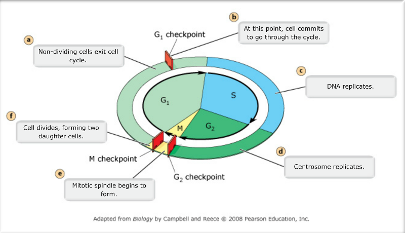
Interphase 90 Cell Cycle Biology Notes Cell Cycle Biology

Anatomy Of The Eye Labeling Eye Anatomy Biology Worksheet Biology Notes

Drag Each Label Into The Proper Position To Identify The Type Of Bone Described In 2022

Cell Membrane Definition Function Structure Animal Plant Cell Cell Membrane Membrane Plasma Membrane


Comments
Post a Comment